Spinal Cord Lesions
Spinal cord lesions are pathologic issues that effect the spinal cord.
Examples of spinal cord lesions include:
- Syringomyelia
- Poliomyelitis
- Werdnig-Hoffman disease
- Amyotrophic lateral sclerosis (ALS)
- Friedrich ataxia
Syringomyelia
Syringomyelia is a cyst in the spinal cord.
Syringomyelias can enlarge, causing spinal compression neurologic symptoms.
Causes of syringomyelia include:
- Cystic degeneration of the spinal cord when there has been trauma
- Arnold-Chiari malformation
Syringomyelia typically occur between C8 and T1.
When the anterior white commissure of the spinothalamic tract is involved a syringomyelia, sensory loss of pain and temperature occurs with sparing of fine touch and position perception in the upper extremities in a “cape like” distribution.
Syringomyelia growth causes other spinal tracts to become involved, which results in weaker, atrophying muscles that have lower muscular tone.
Damage to the lower motor neurons of the anterior horn causes hyporeflexia (diminished reflexes).
 Syringomyelia. Nevit Dilmen. Not altered. CC BY-SA 3.0


Poliomyelitis
Poliomyelitis is due to infection with the poliovirus causes damage to the anterior motor horn.
Lower motor neuron symptoms of poliomyelitis include:
- Flaccid paralysis with atrophying muscles
- Fasciculations
- Weakness with decreased muscle tone
- Poor reflexes
- A negative Babinski sign (down going toes when the bottom of the foot is stroked)
 Poliomyelitis. A child receiving an oral polio vaccine USAID - USAID Bangladesh. Not altered. Public Domain


Werdnig-Hoffman Disease
Werdnig-Hoffman disease is an inherited anterior motor horn degenerative disorder.
Werdnig-Hoffman disease is autosomal recessive.
Werdnig-Hoffman disease presents as a limp, floppy baby that passes away not long after birth.
 Werdnig-Hoffman Disease. Spinal muscular atrophy has an autosomal recessive pattern of inheritance. Domaina, Kashmiri and SUM1 - Not altered. CC BY-SA 3.0

Amytrophic Lateral Sclerosis
Amytrophic lateral sclerosis (ALS) is a corticospinal tract degenerative disease of the upper and lower motor neurons.
Amyotrophic lateral sclerosis is also known as Lou Gehrig disease.
Lower motor neuron signs are caused by anterior motor horn degeneration.
Amyotrophic lateral sclerosis (ALS) lower motor neuron signs include:
- Fasciculations
- Weakness
- Decreased muscle tone
- Poor reflexes
- A negative Babinski sign
- Paralysis
Amyotrophic lateral sclerosis (ALS) upper motor neuron signs include:
- Spastic paralysis
- Hyperreflexia
- Increased muscular tone
- Positive Babinski sign
Amyotrophic lateral sclerosis (ALS) results in lateral corticospinal tract degeneration.
Hand muscle atrophy and weakness are early warning signs of amyotrophic lateral sclerosis (ALS).
The majority of amyotrophic lateral sclerosis (ALS) cases are sporadic and affect middle-aged adults.
Some familial forms of amyotrophic lateral sclerosis are due to zinc-copper superoxide dismutase (SODl) mutation.
The familial zinc-copper superoxide dismutase (SODl) mutation causes free radical damage to neurons, leading to amyotrophic lateral sclerosis.
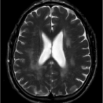
 Amytrophic Lateral Sclerosis. MRI (axial FLAIR) demonstrates increased T2 signal within the posterior part of the internal capsule, consistent with the diagnosis of ALS. Frank Gaillard - Not altered. CC BY-SA 3.0


Friedreich Ataxia
Friedreich ataxia is a disorder that causes spinal cord and cerebellar degeneration.
Friedreich ataxia is brought on by cerebellar degeneration.
Symptoms of Friedreich ataxia include:
- Muscle weakness in the lower limbs
- Loss of deep tendon reflexes
- Loss of vibratory sensation
- Loss of proprioception
Friedreich ataxia is an autosomal recessive condition.
Friedreich ataxia is due to the growth of an unstable trinucleotide repeat (GAA) in the frataxin gene, which causes the frataxin protein to not form or to be abnormally complicated.
Frataxin normally prevents abnormal iron accumulation through it’s effect with mitochondria.
Abnormal or absent frataxin causes an accumulation of iron and free radical damage.
Complications of Friedreich ataxia include:
- Difficulty walking (may need a wheelchair)
- Hypertrophic cardiomyopathy