Vulva
The skin and mucosa of the female genitalia external to the hymen, including the labia majora, labia minora, mons pubis, and vestibule, comprise the vulva.
The vulva is lined by squamous epithelium.
Vulva pathology includes:
- Bartholin cyst
- Condyloma
- Lichen sclerosis
- Lichen simplex chronicus
- Vulvar carcinoma
- Extramammary Paget disease
 Vulva. Labeled image of a vulva, showing external and internal views OpenStax College - Anatomy & Physiology, Connexions Web site. - Not altered. CC BY 3.0

Bartholin’s Gland Cyst
A fluid-filled lump near the vaginal opening is known as a Bartholin’s gland cyst.
Bartholin’s gland cysts can be painful if infected.
Each side of the vaginal canal has one Bartholin’s gland, which produces mucus-like fluid that drains into the lower vestibule via ducts.
Bartholin’s gland cyst develops as a result of inflammation and obstruction of the gland and is most common in women of reproductive age.
Bartholin’s gland cysts appear as a single, painful cystic lesion in the lower vestibule near the vaginal canal.
 Bartholin Cyst. Bartholin's cyst of the right side Medimage. not altered. CC BY-SA 3.0


Condyloma
Condylomas are genital warts.
Condylomas are often large, soft, noncancerous growths that can form on the skin on the outside or inside of the vagina or anus, or inside the cervix.
Condylomas are typically caused by low risk HPV types which include:
- HPV 6
- HPV 11
Histologically, koilocytes, the hallmark of HPV-infected cells, are present in HPV-associated condylomas (technically known as condyloma acuminata).
Condyloma latum is known as secondary syphilis, and is a less frequent cause of condylomas.
Both condyloma acuminatum and condyloma latum are spread sexually.
 Pigmented Seborrehic Keratosis of the Vulva. This was a single SK amid multiple condylomata acuminata removed at the same time. Ed Uthman. Not altered. CC BY 2.0


Lichen Sclerosus
Lichen sclerosus is a localized rash lesion that often itches and is dry.
Lichen sclerosus manifests as a leukoplakia, a white area with vulvar skin that resembles parchment paper.
Postmenopausal women are most frequently affected by this lichen sclerosus, which may have an autoimmune cause.
The histology of lichen sclerosis shows thickening of the epidermis and fibrosis or sclerosus of the dermis are features of lichen sclerosus.
Although lichen sclerosus is regarded as benign, there is an increased risk of squamous cell carcinoma (SCC).
 Lichen Sclerosus, Atrophic. Ed Uthman. Not altered. CC BY 2.0

")

")


Lichen Simplex Chronicus
Lichen simplex chronicus is hyperplasia of the vulvar squamous epithelium.
Lichen simplex chronicus manifests as leukoplakia with thick, leathery vulvar skin and chronic itching and scratching.
Lichen simplex chronicus is regarded as benign.
There is no increased incidence of squamous cell cancer (SCC) with lichen simplex chronicus.
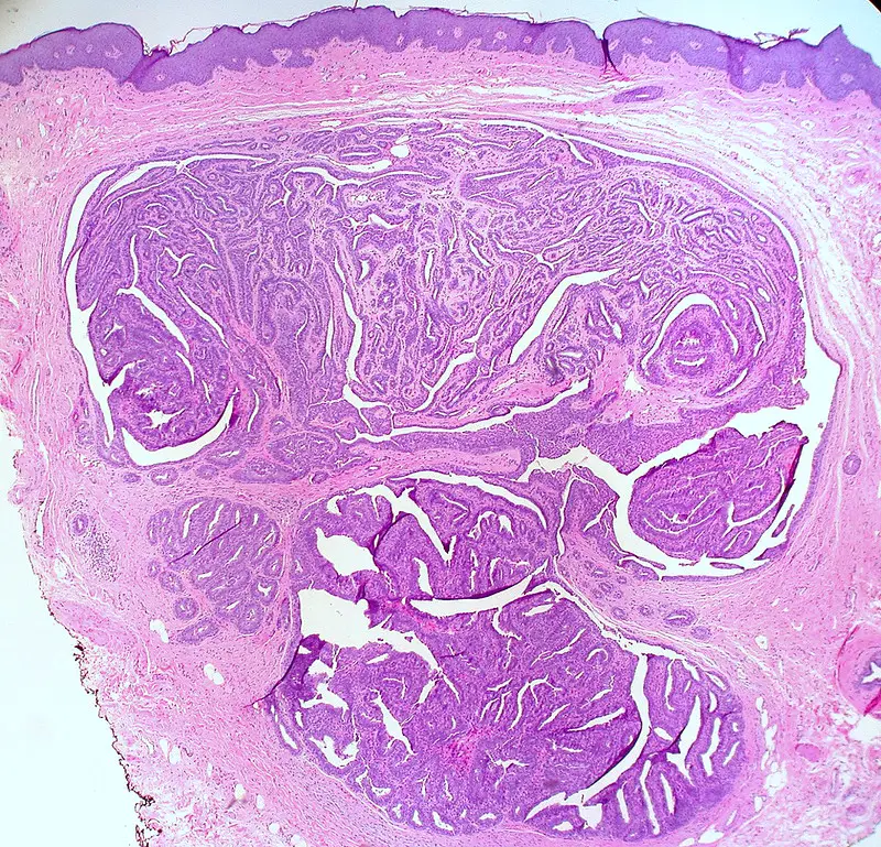
 Lichen Simplex Chronicus. Very low magnification micrograph of lichen simplex chronicus, abbreviated LSC. H&E stain. Skin biopsy. Features: Acanthosis (epidermal thickening). Hyperkeratosis (thickened stratum corneum). Parakeratosis = retention of nuclei in the stratum corneum. +/- Spongiosis (epidermal intercellular edema -- cells appear to have a clear halo around 'em). Nephron. Not altered. CC BY-SA 3.0
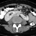
Vulvar Carcinoma
On the external surface of the female genitalia, vulvar carcinoma is a particular form of cancer.
The urethra, vagina, clitoris, and labia are all covered by the vulva, which is the region of skin.
Vulvar carcinoma is rare, making up just a small portion of female genital cancers that manifest as leukoplakia.
The population of females effected by vulvar carcinoma tend to be 70-years-old on average.
To differentiate vulvar carcinoma from other kinds of leukoplakia, a biopsy may be necessary.
The etiology of vulvar carcinoma may be:
- HPV related
- non-HPV related
Risk factors for vulvar carcinoma are:
- Increased number of sexual partners
- Initial age of sexual contact
Vulvar carcinoma develops from vulvar intraepithelial neoplasia, a dysplastic precursor disease characterized histologically by altered koilocytic differentiation, disorganized cellular maturation, nuclear atypia, and elevated mitotic activity.
The most frequent cause of non-HPV associated vulvar cancer is chronic lichen sclerosus.
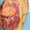
 Vulvar Carcinoma. Stage 4A vulvar cancer Cancer Research UK - Not altered. CC BY-SA 4.0


Extramammary Paget Disease
Extramammary Paget disease is characterized by erythematous, itchy, and ulcerated vulvar skin, which is caused by malignant epithelial cells in the vulva’s epidermis.
In addition to this, extramammary Paget disease also represents carcinoma in situ, usually with no underlying carcinoma.
Note that mammary Paget disease effects the breast.
Extramammary (away from the breast) Paget disease is does not involve the breast.
Melanoma is on the differential for extramammary Paget disease, and special stains must be ordered differentiate the two.
Paget cells are PAS +, keratin +, and S100 -.
Melanoma is PAS -, keratin -, and S100 +.
 Extramammary Paget Disease. H&E stained micrograph of Extramammary Paget's disease, showing Paget cells infiltrating the epidermis Nephron. Not altered. CC BY-SA 3.0