Adrenocortical adenomas are often discovered incidentally and are then termed adrenal incidentalomas (AIs). They are often discovered after an imaging procedure is performed that is unrelated to the adrenal gland.
What is the Pathology of Adrenocortical Adenomas?
The pathology of adrenocortical adenomas is:
-Etiology: The cause of adrenocortical adenomas is associated with chromosomal and genetic abnormalities (genes coding for p53 and p57), the multiple endocrine neoplasia (MEN1) gene is linked to multiple endocrine neoplasia type 1, the aldosynthase/11-beta hydroxylase hybrid gene is associated with glucocorticoid-remediable hyperaldosteronism, and another very rare cause of Cushing syndrome is adrenal-dependent macronodular hyperplasia associated with large adrenal glands.
-Genes involved: p53 and p57, MEN1.
-Pathogenesis: The sequence of events that lead to adrenocortical adenomas depends on the underlying cell type and the cellular mechanisms for primary adrenocortical tumorigenesis are just beginning to be understood.
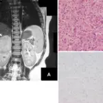
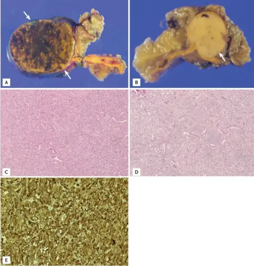
-Morphology: The morphology associated with adrenocortical adenomas shows homogeneous, well-defined, and ovoid mass.
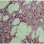
-Histology: The histology associated with adrenocortical adenomas shows increased adrenal tissue.
How do Adrenocortical Adenomas Present?
Patients with adrenocortical adenomas typically are either male or female present at the age range of 70 years or older. The symptoms, features, and clinical findings associated with adrenocortical adenomas include episodic attacks, palpitations, sweats, headaches, and abdominal pain, as well as labile hypertension. Primary hyperaldosteronism includes hypertension. Symptoms also include weight changes, weakness, depression, and bruising.
How are Adrenocortical Adenomas Diagnosed?
Adrenocortical adenomas are diagnosed with biochemical tests.
How are Adrenocortical Adenomas Treated?
Adrenocortical adenomas are treated with surgery for a hormonally active adrenal tumor. The treatment for a malignancy depends on the cell type, spread, and location of the primary tumor and nonfunctional adrenal cortical adenomas are not premalignant, and surgical excision is not indicated.
What is the Prognosis of Adrenocortical Adenomas?
The prognosis of adrenocortical adenomas is good.