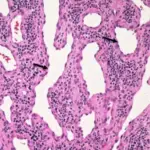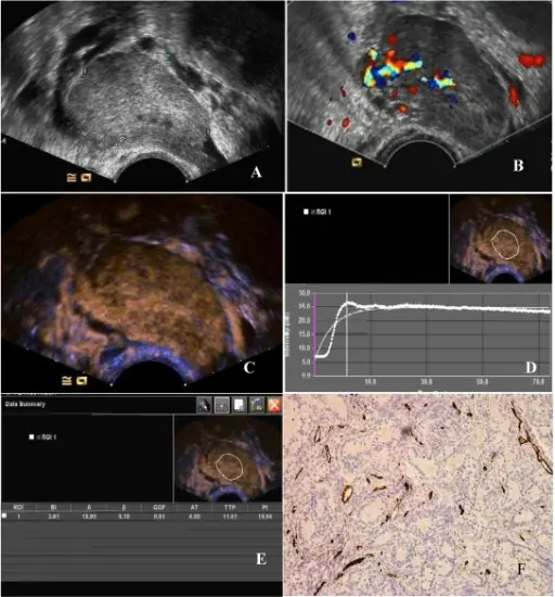
(A) Two-dimensional transvaginal ultrasound showed a hypoechoic mass (4.9 cm × 3.0 cm × 3.3 cm) in the left adnexa with a clear boundary and heterogeneous echo. (B) Abundant blood flow signals can be seen in CDFI. (C) After 11.8 seconds of contrast agent injection, a rapid high enhancement can be seen. (D) A region of interest (ROI) within the tumor was taken to draw a TIC (white curve represents the tumor TIC). (E) The tumor TIC showed that AT was 11.8 seconds, TTP 18.41 seconds, PI is 26.64 dB, AUC 25.95. (F) MVD showed significantly increased interstitial microvessels (CD34, immunohistochemical staining, magnification 200×). Study on the characteristics of contrast-enhanced ultrasound and its utility in assessing the microvessel density in ovarian tumors or tumor-like lesions: Wang J, Lv F, Fei X, Cui Q, Wang L, Gao X, Yuan Z, Lin Q, Lv Y, Liu A - International journal of biological sciences (2011). Not altered. CC.
Benign tumors and tumor-like lesions of the vasculature are neoplasms displaying endothelial cell differentiation, appearing to be resulting from cells that surround blood vessels.
Benign tumors and tumor-Like lesions of the vasculature include:
- Bacillary angiomatosis
- Hemangiomas
- Glomus tumor
- Lymphangiomas
- Vascular ectasias