Brachial cysts are due to a failure of obliteration of the second branchial cleft during embryologic development. Bracial cleft cysts are also known as or cervical lymphoepithelial cysts. Brachial cysts are congenital disorders that form during the fourth week of gestation.
What is the Pathology of Brachial Cysts?
The pathology of branchial cysts is:
-Etiology: The cause of branchial cyst is incomplete involution of branchial cleft structures.
-Genes involved: Unknown.
-Pathogenesis: The internal opening of the branchial sinuses arising from incomplete obliteration in embryogenesis.
-Morphology: They may be on the anterior neck and upper chest bumps.
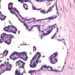
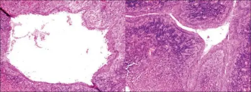
-Histology: The histology associated with branchial cysts shows cysts lined by stratified squamous epithelium that may contain keratinous debris.
How do Branchial Cysts Present?
Patients with branchial cyst typically are equally common in males and females and usually present in childhood or early adulthood. The symptoms, features, and clinical findings associated with branchial cyst include neck lumps.
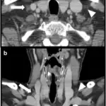
How are Branchial Cysts Diagnosed?
Branchial cysts are diagnosed by ultrasound and biopsy.
How are Branchial Cysts Treated?
Branchial cyst is treated by surgical resection.
What is the Prognosis of Branchial Cysts?
The prognosis of branchial cyst is good.