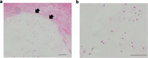
Histology of the resected specimen (H & E stain). (a) Benign hyaline cartilage tumor covered with periosteum. Arrow heads indicate the border between the lesion and the periosteum. (b) Hypocellular cartilage tumor without cytologic atypia and mitosis. Scale bars: 100 μm.A giant periosteal chondroma of the distal femur successfully reconstructed with synthetic bone grafts and a bioresorbable plate: a case report.
Imura Y, Shigi A, Outani H, Hamada K, Tamura H, Morii E, Myoui A, Yoshikawa H, Naka N - World journal of surgical oncology (2014). Not Altered. CC.
Cartilage tumors, also known as chondrogenic tumors, are a type of bone tumor that develops in cartilage and are divided into non-cancerous, cancerous, and intermediate locally aggressive types.
Examples of cartilage-forming tumors include:
- Chordomas
- Osteochondromas
- Chondrosarcomas


