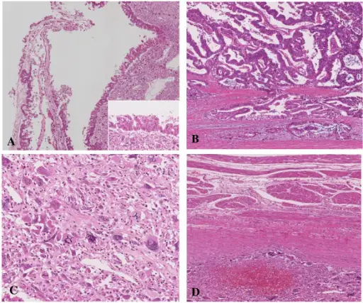
HE staining image of the tumor tissue. (A) The cystic spaces were lined by a columnar mucinous epithelium that presented papillary folding. Higher power view of columnar mucinous epithelium is displayed on the bottom-right corner. (B) There was a small number of stromal invasive features in the bottom of the solid part of this cystic tumor. (C) Near the carcinoma in situ, OGCs were distributed diffusely in the stroma of the cyst wall. (D)The tumor showed the invasion to the stomach across the serosal layer.A male case of an undifferentiated carcinoma with osteoclast-like giant cells originating in an indeterminate mucin-producing cystic neoplasm of the pancreas. A case report and review of the literature. Wada T, Itano O, Oshima G, Chiba N, Ishikawa H, Koyama Y, Du W, Kitagawa Y - World journal of surgical oncology (2011). Not Altered. CC.
Cystic Neoplasms of the pancreas are the fluid-filled sacs within the pancreas.
Examples of Cystic Neoplasms of the pancreas include:
- Intraductal Papillary Mucinous Neoplasms (IPMNs)
- Mucinous Cystic Neoplasms
- Serous Cystic Neoplasms
- Solid-Pseudopapillary Neoplasm


