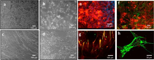
Bright field microscopy images (10×) of (a) HepG2/C3A, (b) iPSC derived human cardiomyocytes, (c) skeletal muscle cells and (d) neurons after 7 days in co-culture in the microfluidic system, in serum free medium and under flow conditions. Immunocytochemical staining of (e) hepatocytes stained for albumin (red) and DAPI (blue), (f) iPSC derived cardiomyocytes stained for troponin (green) and actin (red), (g) skeletal muscle stained for myosin heavy chain (green) and actin (red) and (h) neurons stained for neurofilament (green) and actin (red) after 7 days in co-culture in the system. (scale bars a–g = 100 μm; h = 50 μm).Multi-Organ toxicity demonstration in a functional human in vitro system composed of four organs.
Oleaga C, Bernabini C, Smith AS, Srinivasan B, Jackson M, McLamb W, Platt V, Bridges R, Cai Y, Santhanam N, Berry B, Najjar S, Akanda N, Guo X, Martin C, Ekman G, Esch MB, Langer J, Ouedraogo G, Cotovio J, Breton L, Shuler ML, Hickman JJ - Scientific reports (2016). Not Altered. CC.
Diseases of the skeletal muscle system are diseases that affect the skeletal muscles.
Diseases of the skeletal muscle system include:
- Skeletal muscle atrophy
- Neurogenic and myopathic changes in skeletal muscle
- Inflammatory myopathies
- Toxic myopathies
- Inherited disease of skeletal muscle


