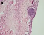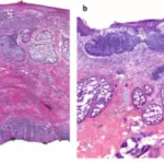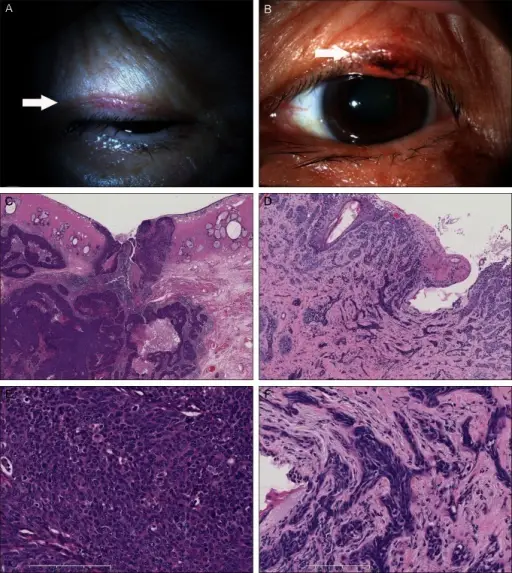
Photographs of eyelid tumors and light microscopic photographs of excised tissues. (A) Photograph of the right eyelid tumor. A small mass is noted (arrow). (B) Photograph of the left eyelid tumor. A slightly elevated lesion with slight erosion is noted at the edge of the eyelid (arrow). (C,E) Light microscope photographs of the right eyelid tumor. Cancer cells proliferate, forming a nest-like structure. Cancer cells have large nuclei and clear cytoplasms (H&E, ×50). (D,F) Light microscope photographs of the left eyelid tumor. Cancer cells proliferate and invade in a morphea-like pattern. Cancer cells have oval-shaped nucleus with hematoxylin-positive cytoplasms (H&E, ×200). A case of Muir-Torre syndrome with multiple cancers of bilateral eyelids and breast.
Kamisasanuki T, Uchino E, Fukushima J, Yoshikawa H, Ishibashi T, Sakamoto T - Korean journal of ophthalmology : KJO (2013). Not Altered. CC.
Eyelid neoplasms are abnormal tissue growth of the eyelid by cellular proliferation that either be benign or malignant. Basal cell carcinoma is the most common malignancy of the eye.
Other neoplasms of the eyelid include sebaceous carcinoma and Kaposi sarcoma.