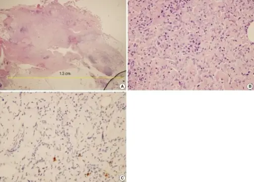
Initial histological appearance of the left breast mass after wide excision. (A, B) Microscopic findings of the specimen showing nodular proliferation of fibrous tissue with focal infiltrating margins. Spindle fibroblasts with many lymphoplasma cells and eosinophils were apparent. A few atypical cells and pleomorphic cells were noted, but abnormal mitosis was not identified (H&E stain; A, ×40; B, ×400). (C) Immunohistochemical staining for S-100 protein shows negative staining in tumors (×200).Undifferentiated pleomorphic sarcoma of the male breast causing diagnostic challenges.
Jeong YJ, Oh HK, Bong JG - Journal of breast cancer (2011). Not Altered. CC.
Fibrous proliferative lesions are characterized by the replacement of normal tissues by a fibrous matrix with various degrees of mineralization and ossification.
Examples of fibrous proliferative lesions include:
- Irritation fibromas
- Peripheral giant cell granulomas
- Pyogenic granulomas
- Peripheral ossifying fibromas


