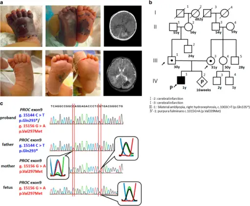
(a) Photograph showing demarcation of healthy and ischemic skin. Necrosis extended to the tendons (arrow) of the right lower leg because of purpura fulminans (PF). PF was completely resolved after anticoagulation therapy (lower left and middle). Computed tomography (CT) image (day 2) and magnetic resonance imaging (MRI, day 3) of the brain, coronal view, showing extensive intracranial thrombosis or hemorrhage in the right occipital lobe. Right upper panel: CT imaging of the brain. Right lower panel: MRI. (b) Familial pedigree. Squares: males; circles: females; open shape: unaffected, noncarrier; half-filled shape: heterozygous carrier; filled shape: congenital protein C deficiency patient. Both parents carried heterozygous mutations in PROC. (c) Prenatal genetic analysis for the fetus. The fetus inherited a wild-type allele from the father and a missense mutation from the mother. Prenatal genetic testing for familial severe congenital protein C deficiency: Tairaku S, Taniguchi-Ikeda M, Okazaki Y, Noguchi Y, Nakamachi Y, Mori T, Kubokawa I, Hayakawa A, Shibata A, Emoto T, Kurahashi H, Toda T, Kawano S, Yamada H, Morioka I, Iijima K - Human genome variation (2015). Not altered. CC.
Diagnostic Methods and Indications for Testing
Laboratory Considerations
Indications for Analysis of Inherited Genetic Alterations
Indications for Analysis of Acquired Genetic Alterations
PCR and Detection of DNA Sequence Alterations
Molecular Analysis of Genomic Alterations
Fluorescence in Situ Hybridization (FISH)
Multiplex Ligation-Dependent Probe Amplification (MLPA)
Southern Blotting
Cytogenomic Array Technology
Polymorphic Markers and Molecular Diagnosis
Polymorphisms and Genome-Wide Analyses
Epigenetic Alterations
RNA Analysis
Next-Generation Sequencing
Bioinformatics
Clinical Applications of NGS DNA
Sequencing
Future Applications