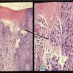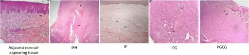
Histological sections representing the normal-appearing oral mucosa and oral reactive lesions (H&E staining ×40). IFH inflammatory fibroepithelial hyperplasia, IF irritation fibroma, PG pyogenic granuloma, PGCG peripheral giant cell lesion. Black arrows indicate normal fibrovascular connective tissue in adjacent oral mucosa, fibrotic connective tissue in IFH, dense collagen bundles in IF, infiltration of inflammatory cells in PG, giant cells in PGCG. Blue arrow indicates endothelium-lined channels in PG. Secretory phospholipase-A2 and fatty acid composition in oral reactive lesions: a cross-sectional study.Ali Hossein Mesgarzadeh H, Akbarzadeh A, Rasipour A, Rasipour T, Mehdizadeh A, Shaaker M.- Cancer Cell Int. (2017). Not Altered. CC.
Inflammatory and Reactive Lesions of the Oral Cavity include:
- Aphthous ulcers
- Fibrous proliferative lesions
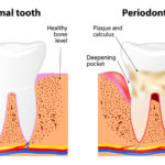 What is Periodontitis?