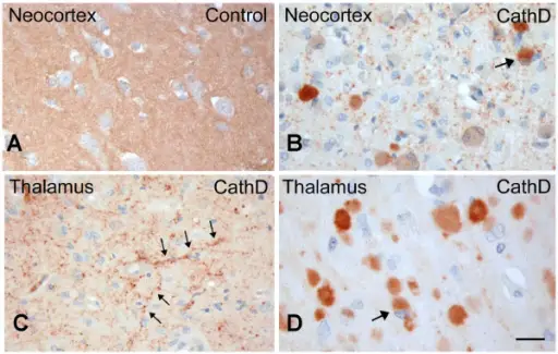
Intracellular accumulations are endogenous by-products and exogenous substances accumulated by injured cells. Examples of intracellular accumulations include:
- Normal cellular constituent accumulated in excess, such as water, lipids, proteins, and carbohydrates
- Abnormal substances (exogenous, such as a mineral or products of infectious agents, or endogenous, such as a product of abnormal synthesis or metabolism)
- Pigments
Intracellular accumulations may occur due to metabolic abnormalities, genetic mutations, or exposure to an indigestible exogenous substance. Some of these accumulations are relatively harmless while others can have drastic effects on health and cognitive function which may include seizures or even death.
What Intracellular Accumulations are due to Lipids?
Intracellular accumulations due to lipids include may result in fatty changes. This is usually apparent in parenchymal cells such as the liver. Steatosis is the accumulation of lipid particles within parenchymal cells. Intracellular lipid accumulation can develop in many organs and tissues, but because the liver is so important to lipid metabolism, hepatic lipidosis is more common.
Atherosclerosis is hardening or thickening of the arteries caused by a buildup of plaque in the inner lining of an artery. Risk factors for atherosclerosis include high cholesterol levels, high triglyceride levels, high blood pressure, smoking, diabetes, obesity, and lack of physical activity.
Cholesterolosis is accumulation of lipid-containing foamy macrophages in the lamina propria of the gallbladder. Grossly this accumulation is seen as yellow mucosal flecks, linear streaks, or a meshlike network.
What Intracellular Accumulations are due to Proteins?
Intracellular accumulations due to proteins are due to the excessive accumulation of aminoacids or proteins that may have negative effects on homeostasis and health.
Examples of intracellular accumulations due to proteins include:
- Alpha-1-antitrypsin deficiency
- Phenylketonuria
What Intracellular Accumulations are due to Hyaline Change?
Intracellular accumulations due to hyaline change include the protein resorption vesicles in the apical cytoplasm of proximal renal tubular epithelial cells that is seen in protein-losing nephropathy. Russell bodies are another example of intracellular accumulations due to hyaline change.
What Intracellular Accumulations are due to Glycogen?
Intracellular accumulations due to glycogen include hepatocellular, clear vacuoles of glycogen in the cytoplasm of cells. They are irregularly shaped than the vacuoles of hepatic lipidosis. They may be seen in glycogen storage diseases.
What Intracellular Accumulations are due to Pigments?
Intracellular accumulations due to pigments include may be from exogenous or endogenous sources.
Exogenous pigments include:
- Carbon (anthracosis)
- Coal dust (pneumoconiosis)
- Tattoo ink (india ink, carbon)
- Ingested (argyria-silver, lead, carotenemia)
Endogenous pigments include:
- Lipofuscin
- Melanin
- Hemosiderin


