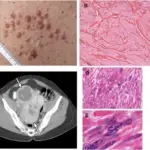
Leiomyosarcomas are rare type of cancer that begins in smooth muscle tissue of uterus.
What is the Pathology of Leiomyosarcomas?
The pathology of leiomyosarcomas is:
-Etiology: The cause of leiomyosarcomas is not exactly known. However, genetic mutation might be one of the causes.
-Genes involved: TP53, RB1, ATRX, PTEN, and MAP2K4 mutations.
-Pathogenesis: The sequence of events that lead to leiomyosarcomas are: Oncogenes control cell growth; tumor suppressor genes control cell division and ensure that cells die at the proper time, abnormal changes in the structure and orientation of these genes result in abnormal growth hence causing leiomyosarcomas.
-Morphology: The morphology associated with leiomyosarcomas shows spindle cell proliferation forming rough bundles and fascicles.
-Histology: The histology associated with leiomyosarcomas shows spindle cells with cigar shaped nuclei with prominent cytologic atypia and mitotic figures.
How does Leiomyosarcomas Present?
Patients with leiomyosarcomas typically in females around age of 50 years. The symptoms, features, and clinical findings associated with leiomyosarcomas include: bloating, fever, pain, nausea, vomiting, weight loss, swelling of skin.
How is Leiomyosarcomas Diagnosed?
Leiomyosarcomas is diagnosed by MRI, CT scan, physical examinations, and biopsy.
How is Leiomyosarcomas Treated?
Leiomyosarcomas is treated by: surgery to remove tumor, radiotherapy, chemotherapy, immunotherapy.
What is the Prognosis of Leiomyosarcomas?
The prognosis of leiomyosarcomas is poor. It is an aggressive cancer that is often diagnosed at later stages, when it has spread to other parts of the body. The 5-year overall survival rate is about 24%.


