Leydig cell tumors are rare testicular growths of the gonadal interstitium that are hormonally active.
What is the Pathology of Leydig Cell Tumors?
The pathology of Leydig cell tumors is:
-Etiology: The cause of Leydig cell tumors is unknown.
-Genes involved: Unknown.
-Pathogenesis: The sequence of events that lead to Leydig cell tumors is unknown though believed endocrine role donates the progress of these tumors.
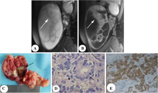
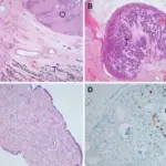
-Morphology: The morphology associated with Leydig cell tumors shows small, well-demarcated, and lobulated nodules.
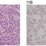
-Histology: The histology associated with Leydig cell tumors shows abundant eosinophilic cytoplasm and Reinke’s crystals.
How does Leydig Cell Tumors Present?
Patients with Leydig cell tumors are typically males that present within an age range of 20 to 50 years old. The symptoms, features, and clinical findings associated with Leydig cell tumors include non-tender profound testicular nodule, gynecomastia, erectile dysfunction, and infertility.
How are Leydig Cell Tumors Diagnosed?
Leydig cell tumors are diagnosed through urine studies, serum studies, and endocrinological tests. Biopsy is confirmatory.
How are Leydig Cell Tumors Treated?
Leydig cell tumors are treated through medical care, chemotherapy, and surgical resection.
What is the Prognosis of Leydig Cell Tumors?
The prognosis of Leydig cell tumors is good for those with benign types, and poor for those with malignant types.


