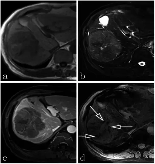
a: no obvious tumor vasculature is visible in the axial T1-weighted imaging or b: the axial T2-weighted imaging; c: the contrast-enhanced axial T1-weighted image shows the mass with irregular enhancement and no obvious tumor veins were detected; d: non-contrast-enhanced SWI shows considerably more detail of the internal architecture than T1-weighted DCE. Scattered linear hypointense signals (arrows) suggest radiating veins in the center of the mass. Characterizing venous vasculatures of hepatocellular carcinoma using a multi-breath-hold two-dimensional susceptibility-weighted imaging: Chang SX, Li GW, Chen Y, Bao H, Zhou L, Yuan J, Wu DM, Dai YM - PloS one (2013). Not altered. CC.
Malignant tumors of the vasculature are tumors that are highly destructive with the potential to recur, and significant distant metastasis.
Malignant tumors of the vasculature include:
- Angiosarcoma
- Hemangiopericytoma
