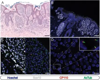
Melanocytic Nevi. Primary cilia are retained by the melanocytes of typical compound and junctional cutaneous melanocytic nevi.Immunostaining as indicated. (A and B) Low-power images of cutaneous melanocytic nevi stained with hematoxylin and eosin (A) or for immunofluorescence microscopy (B). Scale bars: 100 µm. (C) Split image of nested melanocytes within a melanocytic nevus. Sox10 epifluorescence has been omitted from the right half of the image to enhance visualization of cilia and centriole staining. Scale bar: 50 µm. (D) High power image of nest shown in panel C. Scale bar: 25 µm. Primary cilium depletion typifies cutaneous melanoma in situ and malignant melanoma: Kim J, Dabiri S, Seeley ES - PloS one (2011). Not altered. CC.
Melanocytic nevi are the cancer of the melanocytes either congenital or acquired.
Examples of melanocytic nevi include:
- Congenital nevus
- Blue nevus
- Spitz nevus
- Halo nevus
- Dysplastic nevus
 What is Lentigo?