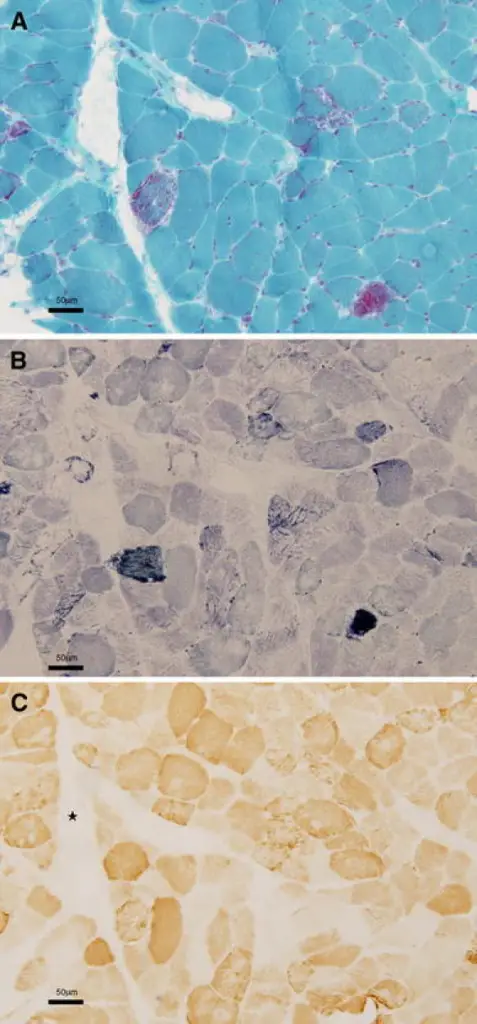
Light microscopy of muscle biopsy from patient II-4. a Modified Gomori trichrome staining shows several ragged red fibers. b Succinate dehydrogenase reaction (SDH) shows numerous hyperintense fibers. Strongly SDH-reactive blood vessel (SSV) is highlighted. c Cytochrome c oxidase (COX) reaction shows several negative fibers. Asterisk indicates that SSV is COX-negative. Scale bars represent 50 µm (color figure online)ariable phenotypes in a family with mitochondrial encephalomyopathy harboring a 3291T > C mutation in mitochondrial DNA.
Sunami Y, Sugaya K, Chihara N, Goto Y, Matsubara S - Neurological sciences : official journal of the Italian Neurological Society and of the Italian Society of Clinical Neurophysiology (2011). Not Altered. CC.
Mitochondrial encephalomyopathies represent a clinically heterogeneous group of disorders resulting from abnormal mitochondrial function. Organs with high mitochondrial loads (like the brain and muscles) are particularly susceptible to mitochondrial dysfunction.
Examples of mitochondrial encephalomyopathies include:
- Leigh syndrome
- Mitochondrial encephalomyopathy lactic acidosis and stroke-like episodes (MELAS)
- Myoclonic epilepsy and ragged red fibers (MERRF)


