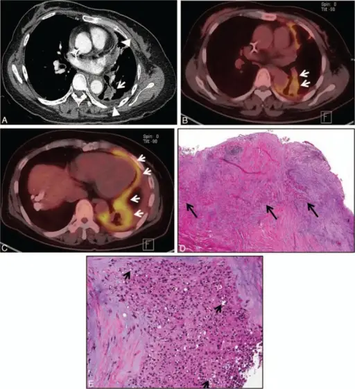
Epithelioid hemangioendothelioma of parenchymal tumor(s) with pleural extension pattern in a 53-y-old woman. (A) Mediastinal window images of enhanced CT scan demonstrate subpleural lung tumor (arrow) and tumor pleural seeding (arrowheads). (B and C) PET/CT images obtained at the levels of right inferior pulmonary vein (B) and liver dome (C) depict increased FDG uptake in lung (arrows in B) and pleural (arrows in C) lesions. (D) Low-magnification photomicrograph of parietal (diaphragmatic) pleural biopsy specimen depicts thickened pleura containing areas of tumor cells (arrows). (E) High-magnification photomicrograph reveals epithelioid tumor cells containing prominent cytoplasmic vacuoles or intracytoplasmic lumina (arrows). CT = computed tomography, FDG = fluorodeoxyglucose, PET = positron emission tomography.Epithelioid hemangioendothelioma in the thorax: Clinicopathologic, CT, PET, and prognostic features
Medicine. Not Altered. CC.
Examples of other parenchymal tumors include:
- Primary CNS lymphoma
- Germ cell tumors
- Pineal parenchymal tumors.
 What is an Atypical Rhabdoid Tumor?