Peripheral B-cell neoplasms are neoplasms involving mature B-cell lineage.
Examples of peripheral B-cell neoplasms include:
- Chronic lymphocytic leukemia/small lymphocytic lymphoma
- B-cell prolymphocytic leukemia
- Lymphoplasmacytic lymphoma
- Splenic marginal zone lymphoma
- Nodal marginal zone lymphomas
- Extranodal marginal zone lymphoma
- Mantle cell lymphoma
- Follicular lymphoma
- Marginal zone lymphoma
- Hairy cell leukemia
- Diffuse large B-cell lymphoma
- Burkitt lymphoma
- Plasma cell neoplasms and related conditions
| Peripheral B-cell neoplasm | Genetics | Key Histologic Features | Key Clinical Findings |
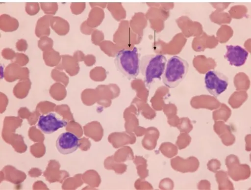
| Chronic lymphocytic leukemia/small lymphocytic lymphoma | Deletions of 13q14.3, 11q, 17p and trisomy 12q | Small lymphocytes with condensed chromatin and scant cytoplasm. Smudge cells, which are disrupted tumor cells. Larger activated lymphocytes that often gather in loose aggregates are referred to as proliferation centers, which are pathognomonic for CLL/SLL | Easy fatigability, weight loss and anorexia, generalized lymphadenopathy, hepatosplenomegaly |
| B-cell prolymphocytic leukemia | TP53 gene, MYC gene | Large lymphoid cells (prolymphocytes) accounting for at least 55% of total circulating cells in the peripheral blood | High lymphocyte count, splenomegaly, B-symptoms (fevers, night sweats, weight loss), anemia and thrombocytopenia |
| Splenic marginal zone lymphoma | TP53 gene, NOTCH2 gene, KLF2 gene | Prominent expansion of the white pulp by small lymphocytes that often replace the normal germinal centers and efface the normal mantle zones | Massive splenomegaly, autoimmune anemia, thrombocytopenia, neutropenia, absolute lymphocytosis |
| Nodal marginal zone lymphoma | Ig gene | Perifollicular and interfollicular lymphoid infiltrate that surrounds variably preserved germinal centers; diffuse pattern of infiltration | Unexplained fever, drenching night sweats and unexplained weight loss, localized or generalized peripheral lymphadenopathy. |
| Extranodal marginal zone lymphoma | MALT1 gene, BCL10 gene | Poorly defined follicular appearing areas that are composed of monocytoid B cells that feature enlarged nuclei | Fever without an infection, night sweats, unexplained weight loss, skin rash, chest or abdominal pain, tiredness. |
| Mantle cell lymphoma | ATM, CCND1, TP53, MLL2, TRAF2 and NOTCH1 genes | Expansion of the mantle zone that surrounds the lymph node germinal centers by small-to-medium atypical lymphocytes | Fever, night sweats, weight loss, generalized lymphadenopathy, abdominal distention from hepatosplenomegaly, fatigue from anemia or bulky disease. |
| Follicular lymphoma | KMT2D gene | Nodular and diffuse growth pattern in involved lymph nodes. Two cell types are present: centrocytes and centroblasts | Painless, generalized lymphadenopathy |
| Marginal zone lymphoma | NOTCH gene, KLF2 gene, PTPRD gene | Small to medium sized lymphocytes surrounding a reactive follicle. Plasmacytic differentiation is common. | Fever without an infection, night sweats, unexplained weight loss, skin rash, chest or abdominal pain and tiredness |
| Hairy cell leukemia | BRAF serine/threonine kinase gene | Diffuse interstitial infiltrate of cells with round, oblong or reniform nuclei and moderate amounts of pale blue cytoplasm with thread-like or bleb-like extensions | Massive splenomegaly is the most common and sometimes the only abnormal physical finding |
| Diffuse large B-cell lymphoma | BCL6, BCL2, p300 and CREBP genes | Relatively large cell size usually 4-5 times the diameter of a small lymphocyte and a diffuse pattern of growth | Rapidly enlarging mass at a nodal or extranodal site. The Waldeyer ring, oropharyngeal lymphoid tissue that includes the tonsils and adenoids, are most commonly involved. |
| Burkitt lymphoma | MYC gene | Diffuse infiltrate of intermediate-sized lymphoid cells 10 to 25 um in diameter with round or oval nuclei, coarse chromatin, several nucleoli and moderate amount of cytoplasm. | Endemic Burkitt lymphoma often presents as a mass involving the mandible and shows an unusual predilection for involvement of abdominal viscera (kidney, ovaries and adrenal glands). Sporadic Burkitt lymphoma most often appears as a mass involving the ileocecum and peritoneum. |


