Pinealomas are fairly uncommon type of brain tumor mostly encountered in childhood. The pineal gland is part of the epithalamus, a posterior segment of diencephalon, that secrets melatonin and is responsible for circadian rhythms. Pineal tumors and cysts may cause precocious puberty and delayed puberty.
What is the Pathology of Pinealomas?
The pathology of pinealomas is:
-Etiology: The cause of pinealomas is unknown. The tumor may be caused by genetic predisposition or environmental influence.
-Genes involved: Mutations to the RB1 gene.
-Pathogenesis: Proliferation of abberent germinal cells.
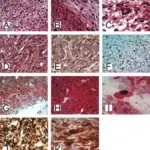
-Histology: The histology associated with pinealoma depends on which type of tumor that grows from pineocytoma.
How does Pinealoma Present?
Patients with a pinealoma typically could have this tumor also can cause hydrocephalus, gait disturbances or precocious puberty.
How is Pinealoma Diagnosed?
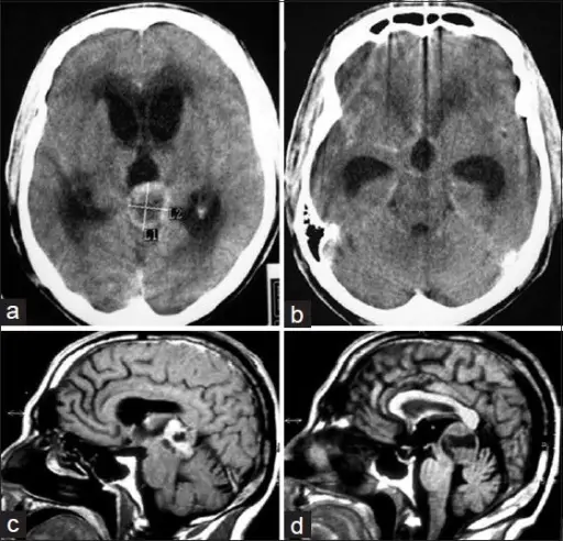
Pinealoma is diagnosed through several methods, which are central and peripheral vision assessment, opthalmosocpic assessment to check for optic nerve swelling which can signify increased intracranial pressure. Head MRI scans and CT scans to determine the size, shape, and location of the pinealoma. A biopsy allows determine the type and grade of the tumor.
How is Pinealoma Treated?
Surgical removal which may be followed by radiation therapy, chemotherapy, targeted therapy, or immunotherapy.
What is the Prognosis of Pinealoma?
The prognosis of pinealoma depends on type and grade of tumor, patient`s age and gender and how the disease responds to treatment. The relative 5-year survival rate for pineal region tumors is 70%.