Pneumoconioses are a group of interstitial lung diseases usually caused by inhalation of dust, mostly at work.
What is the Pathology of Pneumoconioses?
The pathology of pneumoconioses is:
-Etiology: The cause of pneumoconioses is occupational exposure to specific airborne agents.
-Genes involved: None.
-Pathogenesis: The sequence of events that lead to pneumoconioses is as follows inhaled particles deposited at the distal airways and alveoli. There is phagocytizing of the particles by macrophages. profibrotic pathways and proinflammatory are activated. Consequent exposure and ingestion of dust by macrophages causes cell necrosis and autophagy, repeated cycles cause advanced alveolar fibrosis and inflammation.
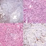
-Histology: The histology associated with pneumoconioses shows aggregates of dust-laden macrophages at the bronchiolar and alveolar walls. Distended bronchioles and alveoli surrounding the macules.
Table of Common Pneumoconiosis
| Agents | Diseases |
| Coal | Simple coal-workers’ pneumoconiosisProgressive massive fibrosisCaplan’s syndrome |
| Silica | SilicosisCaplan’s syndrome |
| Asbestos | Asbestosis Pleural diseasesTumors |
| Beryllium | Berylliosis |
| Mouldy hay | Farmer’s lung |
How does Pneumoconiosis Present?
Patients with pneumoconiosis typically have varying preferences male workers affected most. present at age range of above 45 years. The symptoms, features, and clinical findings associated with pneumoconiosis include; exercise tolerance reduction, the steady beginning of a cough nonproductive, some are symptomless with an anomalous chest X-ray.
How is Pneumoconiosis Diagnosed?
Pneumoconiosis is diagnosed through a history of long-time exposure to agents. Pulmonary function tests show a characteristic restrictive outline. Imaging studies- chest X-ray, CT scan showing small diffuse nodules.
How is Pneumoconiosis Treated?
Pneumoconiosis is treated through enhanced workout tolerance and avoidance of causative exposure. There is no cure.
What is the Prognosis of Pneumoconiosis?
The prognosis of pneumoconiosis is poor in a fibrotic stage of illnesses.