Proteosomes are protein complexes that degrade unnecessary or damaged proteins by proteolysis.
What are Proteosomes?

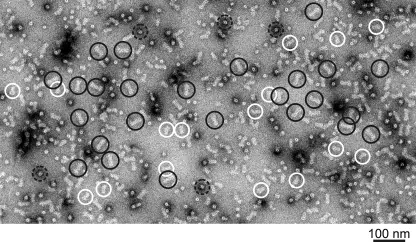
Proteosomes. Electron micrograph of a negatively stained 26S proteasome sample. Examples of side views of double-capped and single-capped 26S proteasome images are identified with black and white circles, respectively. End views of 26S proteasome complexes are identified by black dashed circles. Structure of the human 26S proteasome: subunit radial displacements open the gate into the proteolytic core. da Fonseca PC, Morris EP - The Journal of biological chemistry (2008). Not Altered. CC.


