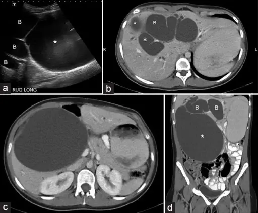
(a) Ultrasound scan axial view shows a large cystic mass (*) and dilated bile ducts (B). The mass was very large and the relationship of the biliary tree to the mass was not clear on ultrasound. (b) Computed tomography (CT) scan, axial view of portal venous phase demonstrates dilated intrahepatic ducts (B). The superior portion of the cystic mass is visualized (*). (c) Computed tomography scan axial view at a lower level than 1b shows a large cystic mass (*) at the porta hepatis. (d) Coronal reformatted computed tomography image delineates a huge cystic mass in the right upper quadrant (*). Dilated bile ducts are visualized (B). Giant choledochal cyst mimicking massive gallbladder hydrops in an adult patient: multidetector computed tomography and magnetic resonance imaging findings correlated to gross and histopathological findings: Choi JI, Lall C, Bhargava P, Imagawa DK - Journal of clinical imaging science (2013). Not altered. CC.
Structural anomalies of the biliary tree are the anomalies of the pancreatic or bile duct.
Examples of structural anomalies of the biliary tree include:
- Choledochal cysts
- Fibropolycystic disease
