Teratomas are complex tumors having several cellular or organoid components suggestive of normal derivatives from more than one germ layer.
What is the Pathology of Teratomas?
The pathology of teratomas is:
-Etiology: The cause of teratomas is associated with prenatal risk factors such as low birth weight, cryptorchidism, and genetic predisposition.
-Genes involved: metalloproteinase MMP7 gene, anti-apoptotic EGR1 gene.
-Pathogenesis: The sequence of events that lead to teratomas; metamorphoses in the genes linked to membrane-membrane interface, matrix establishment, and remodeling enzymes.
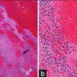
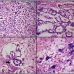
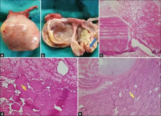
-Morphology: The morphology associated with teratomas shows characteristic presence of hair, bones, teeth, cartilage, and cystic spaces.
-Histology: The histology associated with teratomas shows a heterogeneous, collection of distinguished cells or organoid structures.
How does Teratomas Present?
Patients with teratomas typically found in male children and young adults, present at an age range of 20 to 35 years. The symptoms, features, and clinical findings associated with teratomas include palpable masses.
How are Teratomas Diagnosed?
Teratomas are diagnosed through laboratory studies serological tests, serum tumor makers. Imaging studies retroperitoneal/abdominal ultrasound and CT scan, X-rays, and biopsies.
How is Teratomas Treated?
Teratomas are treated through medical care- cisplatin chemotherapy.
What is the Prognosis of Teratomas?
The prognosis of teratomas is fairly grounded on classification with a mortality rate of 10% of good, 30% of intermediate, and 60% of poor.