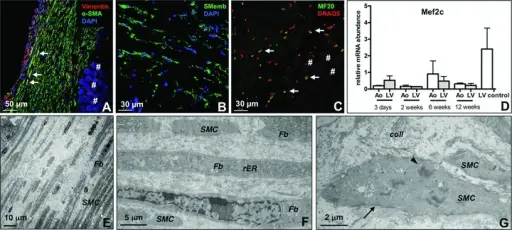
Interactions with the Extracellular Matrix. (A) Fibroblasts are stained with vimentin (red), smooth muscle cells are stained with a-smooth muscle actin (α-SMA, green). Colocalization of vimentin and α-SMA is typical for myofibroblasts, which appear yellow (arrows). Nuclei are blue (#: implanted membrane). (B) Myofibroblasts specifically stained with anti-non-muscle myosin heavy chain b (SMemb; green); nuclei are blue. (C) Cells expressing myosin heavy chain were stained with MF20 (arrows, green), nuclei are red (#: implanted membrane). (D) Mef2c, a marker of muscle cells, was detected in aortic (Ao) and left ventricular membranes (qRT-PCR; n = 8 per time-point). (E) Electron microscopy image showing clear orientation of cells in the newly developed tissue after 12 weeks. (F) Electron microscopy image showing different types of differentiated cells after 6 weeks (SMC, smooth muscle cell; Fb, fibroblast; rER, rough endoplasmic reticulum). (G) Electron microscopy image showing SMCs and organized collagen fibres (coll). Intracellular dense bodies (arrowhead) indicate terminal differentiation and contractile function of SMCs. Focal adhesion points (arrow) hallmark cellular interaction with the extracellular matrix, implying functional organization. Stem cell-mediated natural tissue engineering. Möllmann H, Nef HM, Voss S, Troidl C, Willmer M, Szardien S, Rolf A, Klement M, Voswinckel R, Kostin S, Ghofrani HA, Hamm CW, Elsässer A - Journal of cellular and molecular medicine (2011). Not Altered. CC.
Interactions with the extracellular matrix include:
- Basement membrane foundation
- Mechanical support
- Scaffolding for tissue renewal
There are two basic forms of the extracellular matrix:
- Basement membrane
- Interstitial matrix
Components of the extracellular matrix include:
- Fibrous structural proteins like elastin and collagen
- Adhesive glycoproteins that aid with connectivity
- Water-hydrated gels like hyaluronic acid and proteoglycans


