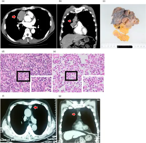
(a,b) Computed tomography (CT) findings of thymomas of Patient 1. (a) Horizontal plane, (b) Coronal plane. Arrows indicate thymoma. (c) The macroscopic finding of the thymoma in patient 1. (d,e) Hematoxylin and eosin stain of thymoma tissues. (d) Patient 1, (e) Patient 2 (×400). (f,g) CT findings of the thymoma of patient 3. (f) Horizontal plane, (g) Coronal plane. Arrows indicate thymoma. A novel thymoma-associated autoimmune disease: Anti-PIT-1 antibody syndrome: Scientific Reports. Not altered. CC.
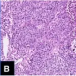
Thymomas are tumors of thymic epithelial cell origin.
The types of thymomas are:
- Type A thymoma, including atypical variant
- Type AB thymoma
- Type B1 thymoma
- Type B2 thymoma
- Type B3 thymoma
- Micronodular thymoma with lymphoid stroma
- Metaplastic thymoma
- Microscopic thymoma
- Sclerosing thymoma
- Lipofibroadenoma
Useful immunohistochemistry includes: AE1/AE3 (pan-keratins), CK19, CK20, CD5, CD20, CD117, TdT, p40, p63,


