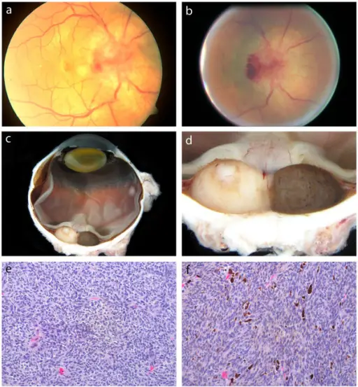
Clinical and pathological findings. (a) Initial dilated fundus examination: disc edema, hemorrhage and a serous retinal detachment. (b) Fundus examination 10 months later: increase in the size of the mass, with disc border hemorrhage and an adjacent ring of pigmentation. (c) (d) Gross pathology: uveal melanoma with distinct non-pigmented and pigmented halves. (e) Histopathological section of the non-pigmented half of the uveal melanoma, spindle cell type. (f) Histopathological section of the pigmented half of the uveal melanoma, of spindle cell type with melanin. Case report: an atypical peripapillary uveal melanoma. Lim LA, Miyamoto C, Blanco P, Bakalian S, Burnier MN - BMC ophthalmology (2014). Not Altered. CC.
Uveal melanomas are cancers of the eye involving the tissues of the uvea.
What is the Pathology of Uveal Melanoma?
The pathology of uveal melanoma is malignant proliferation of melanocytes in the uvea.
How does Uveal Melanoma Present?
Uveal melanoma presents with proptosis, photopsia, diplosia, metamorphopsia and may be asymptomatic in some cases.
How is Uveal Melanoma Diagnosed?
Uveal melanoma is diagnosed by ophthalmoscopy, and biomicroscopy.
How is Uveal Melanoma Treated?
Uveal melanoma is treated with photocoagulation photodynamic therapy, proton beam radiotherapy, transpupillary thermotherapy, and brachytherapy enucleation.
What is the Prognosis of Uveal Melanoma?
The prognosis of uveal melanoma is poor.