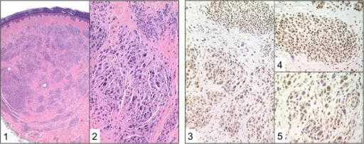
Histology and immunohistochemistry of MBAITs. Representative biopsy from individual W-III-04. The histologic examination (hematoxylin and eosin, H/E, of the nevi at the low power (H&E, 4X Original Magnification) magnification shows an intradermal melanocytic nevus with superficial nests and deeper single melanocytic units. This background melanocytic nevus shows maturation with depth. A second distinct, central, circumscribed population of melanocytes is identified at the mid-reticular dermis (1). This second population of melanocytes on higher power (H&E, 20X Original Magnification) shows variably sized, large epithelioid and spindled melanocytes. There is marked cytologic atypia comprised of pleomorphic, hyperchromatic nuclei (2). The immunohistochemistry for BAP1 demonstrates distinct staining between the background melanocytic nevus and the embedded clusters of atypical epithelioid melanocytes. Identified is strong nuclear positivity in the background melanocytic nevus and negative nuclear staining in the large epithelioid cells (3: BAP-1, 20X Original Magnification). On higher power, the background melanocytes show strong nuclear staining in this superficial nest (4: BAP-1, 40X Original Magnification). Large epithelioid cells with their pleomorphic nuclei demonstrate negative nuclear staining with variable cytoplasmic staining (5: BAP-1, 40X Original Magnification).BAP1 cancer syndrome: malignant mesothelioma, uveal and cutaneous melanoma, and MBAITs.
Carbone M, Ferris LK, Baumann F, Napolitano A, Lum CA, Flores EG, Gaudino G, Powers A, Bryant-Greenwood P, Krausz T, Hyjek E, Tate R, Friedberg J, Weigel T, Pass HI, Yang H - Journal of translational medicine (2012). Not Altered. CC.
