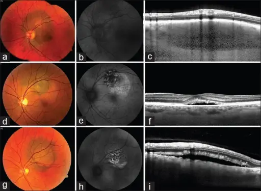
Clinical and imaging features of choroidal melanocytic lesions. Case 1: Circumpapillary pigmented choroidal lesion lacking orange pigment (a). Fundus autoflouroscense (b) is indistinct with no lipofuscin. Enhanced depth imaging-optical coherence tomography (c) normal photoreceptors and absence of SRF, consistent with choroidal nevus. Case 2: Pigmented choroidal lesion with overlying orange pigment, diffuse hyperautofluorescence (e) and the subfoveal fluid with shaggy photoreceptors (f) suggestive of choroidal melanoma. Case 3: Pigmented choroidal lesion with orange pigment (g), patchy hyperautofluorescence with sedimentation (h), and overlying SRF and shaggy photoreceptors (i), suggestive of choroidal
melanoma. Using risk factors for detection and prognostication of uveal melanoma. Rishi P, Koundanya VV, Shields CL - Indian journal of ophthalmology (2015). Not Altered. CC.
Uveal nevi are benign lesions of the uvea layer of the eye. Uvea nevi rarely produces glaucoma.
What is the Pathology of Uveal Nevi?
The pathology of uveal nevi is moles of unclear deposits on the eyeball and other hyperpigmentation spots.
How does Uveal Nevi Present?
Uveal nevi presents with visible freckles in the eye.
How is Uveal Nevi Diagnosed?
Uveal nevi is diagnosed by fundoscopy.
How is Uveal Nevi Treated?
Uveal nevi is treated with local excision, or argon laser photoablation.
What is the Prognosis of Uveal Nevi?
The prognosis of uveal nevi is typically fine.