Cardiac lipoma is an encapsulated, benign, primary cardiac tumor considered to be the most common non-myxomatous cardiac tumor.
What is the Pathology of Cardiac Lipoma?
The pathology of cardiac lipoma is:
-Etiology: The cause of cardiac lipoma is unknown, however, association with gene rearrangement is seen.
-Genes involved: chromosome 12 gene rearrangement, HMGA2-LPP fusion.
-Pathogenesis: The sequence of events that lead to cardiac lipoma is not understood but to progression of the tumor may be silent for a long period of time and may grow in size without infiltrating the myocardium. It may result in myocardium resorption and cavitation that may involve the cardiac chambers.
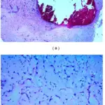
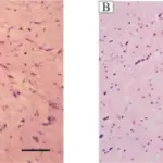
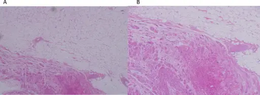
-Histology: The histology associated with cardiac lipoma shows mature fat cells within the collagenous capsule.
How does Cardiac Lipoma Present?
Patients with cardiac lipoma have no sex or age predilection. The symptoms, features, and clinical findings associated with cardiac lipoma include dyspnea, syncope, arrhythmia, palpitations, and angina.
How is Cardiac Lipoma Diagnosed?
Cardiac lipoma is diagnosed in echocardiography showing a cardiac mass. CT and MRI are good modalities to show tissue characteristics; coronary arteriography can aid the operating surgeon in outlining blood supply and coronary anatomy.
How is Cardiac Lipoma Treated?
Cardiac lipoma is treated through surgical excision of the tumor. However, treatment is not warranted in asymptomatic individuals.
What is the Prognosis of Cardiac Lipoma?
The prognosis of cardiac lipoma is good with patients not usually needing treatment or surgical intervention, but if needed, resection can be done easily followed with excellent patient recovery.