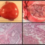
A complete mole is the most common type and does not contain metal parts.
What is the Pathology of Complete Mole?
The pathology of the complete mole is:
-Etiology: The cause of a complete mole is a single sperm (90% of the time) or two (10% of the time) sperm combining with an egg that has lost its DNA.
-Genes involved: None.
-Pathogenesis: The sequence of events that lead to complete mole shows diploid (45, XX, rarely 46, XY), absent fetus/embryo, diffuse swelling of villi, and diffuse trophoblastic hyperplasia.
-Morphology: The morphology associated with complete mole shows all the chorionic villi are vesicular, and no sign of embryonic or fetal development is present.
-Histology: The histology associated with complete mole shows diffuse, villous enlargement with marked hydropic changes.
How does Complete Mole Present?
Patients with complete mole typically affect females present at the age range of 18-55 years. The symptoms, features, and clinical findings associated with complete mole include painless vaginal bleeding, the uterus may be larger than expected, or the ovaries may be enlarged, hyperemesis, increased blood pressure, high HCG levels.
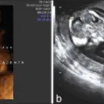
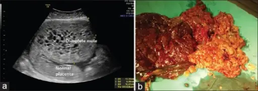
How is Complete Mole Diagnosed?
Complete mole is diagnosed using ultrasound and histopathological examination.
How is Complete Mole Treated?
Complete mole is treated by uterine suction or by surgical curettage, and methotrexate.
What is the Prognosis of Complete Mole?
The prognosis of the complete mole is good if treated in a timely manner.