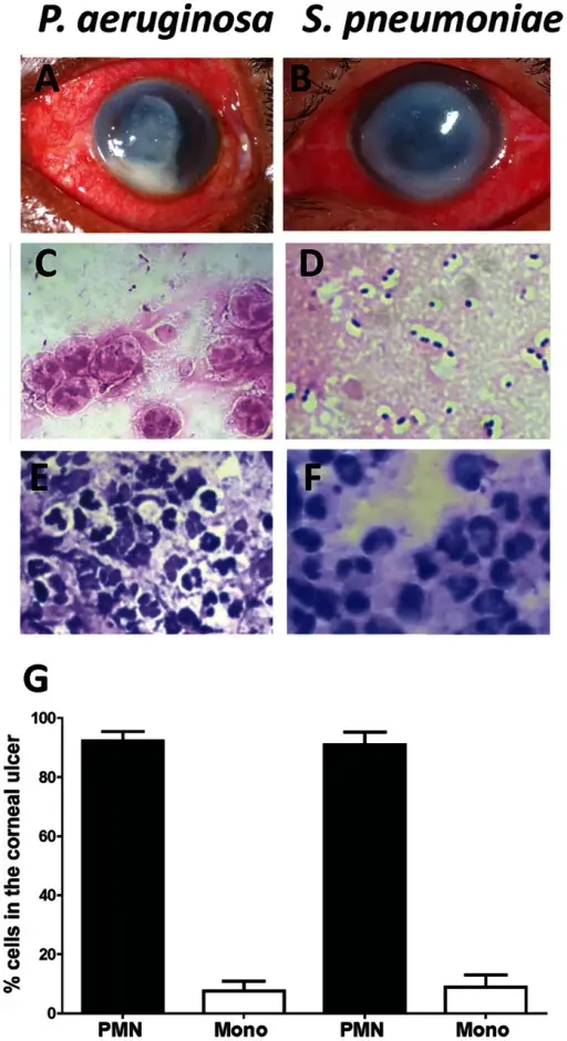
Cellular composition of corneal ulcers from patients with bacterial keratitis.Representative corneal ulcers of patients caused by P. aeruginosa (A), or by S. pneumoniae (B). C, D. Gram staining of corneal ulcer material showing Gram negative bacilli (C), and Gram positive diplococci and chains (D). Original magnification is x1000. E,F: Wrights Giemsa (Diff-Quik) stain of corneal ulcer material from P. aeruginosa (E), or S. pneumoniae (F) infected tissue Original magnification is x400. G. Percent neutrophils and mononuclear cells were determined by counting cells from ten P. aeruginosa and ten S. pneumoniae patients.Host response and bacterial virulence factor expression in Pseudomonas aeruginosa and Streptococcus pneumoniae corneal ulcers.
Karthikeyan RS, Priya JL, Leal SM, Toska J, Rietsch A, Prajna V, Pearlman E, Lalitha P - PloS one (2013). Not Altered. CC.
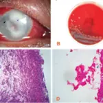
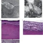
Corneal ulcers are loss of tissue of the cornea resulting in open soreness.
What is the Pathology of Corneal Ulcer?
The pathology of corneal ulcer is vitamin A deficiency, fungal, viral and amoebic infection of the cornea.
How does Corneal Ulcer Present?
Corneal ulcer presents with itchy eyes, pus-like discharge from the eyes, and watery eyes.
How is Corneal Ulcer Diagnosed?
Corneal ulcer is diagnosed by fluorescein eye stain, histological examination, etc.
How is Corneal Ulcer Treated?
Corneal ulcer is treated with antibacterial, antifungal, and antiviral eye drops and medications, and corneal transplant.
What is the Prognosis of Corneal Ulcer?
The prognosis of corneal ulcer is the patient may respond to treatment but may lead to partial loss of vision.