Dermatofibroma is a nodule of cutaneous origin and is also known as the cutaneous fibrous histiocytoma.
What is the Pathology of Dermatofibroma?
The pathology of dermatofibroma is the scientific study of the dermis and the conditions arising from the dermis.
-Etiology: The cause of dermatofibroma can be trauma e.g. insect bite, change in immunity, and genetic predisposition.
-Genes involved: None.
-Pathogenesis: The sequence of events that lead to dermatofibroma is the continuous excessive growth of the fibrous tissue of the dermis, is not known.
-Morphology: The morphology associated with dermatofibroma shows painless pink firm nodule with some itchiness and may vary in color as time goes by.
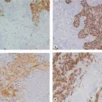
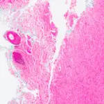
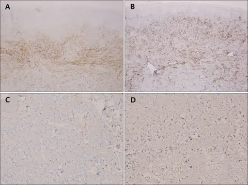
-Histology: The histology associated with dermatofibroma shows acanthotic epithelium with basilar hyperpigmentation and collagen trapping by dermal fibrohystiocytic infiltrates and are well circumscribed.
How does Dermatofibroma Present?
Patients with dermatofibroma typically occur in females and present at the age range of teenagers to adults. The symptoms, features, and clinical findings associated with dermatofibroma include itchiness and tenderness but are painless redness.
How is Dermatofibroma Diagnosed?
The dermatofibroma is diagnosed by physical examination, biopsy, and sonography.
How is Dermatofibroma Treated?
The dermatofibroma is treated by cryotherapy, steroid injection, surgical excision.
What is the Prognosis of Dermatofibroma?
The prognosis of dermatofibroma is good since they are slow-growing and are noncancerous.