A germinal matrix hemorrhage is a bleeding into the subependymal germinal matrix. There may be subsequent rupture into the lateral ventricle.
What is the Pathology of Germinal Matrix Hemorrhage?
The pathology of germinal matrix hemorrhage is:
–Etiology: The cause of germinal matrix hemorrhage is the weak blood vessels of the germinal matrix are predisposed to hemorrhage. Stress experienced by a premature baby after birth may cause these weak vessels burst. The subsequent bleeding occurs initially in the periventricular areas causing a periventricular hemorrhage.
–Genes involved: Methylenetetrahydrofolate reductase variants
-Pathogenesis: The sequence of events that lead to germinal matrix hemorrhage are fragile thin-walled blood vessels that rupture. Hence the microcirculation in this particular area is extremely sensitive to hypoxia and changes in perfusion pressure. It is most frequent before 35 weeks gestation and is typically seen in very low birth-weight premature infants due to their decreased ability for regulate cerebral blood flow. Increased arterial blood pressure in these blood vessels leads to rupture and hemorrhage into the germinal matrix.
–Morphologic changes: The morphologic changes involved with germinal matrix hemorrhage are pooling of blood in a potential space.
How does Germinal Matrix Hemorrhage Present?
Patients with germinal matrix hemorrhage typically have no gender preference. Premature infants are at increased risk. The symptoms, features, and clinical findings associated with germinal matrix hemorrhage includes nose bleeds, respiratory depression, abnormal posturing, enlarged head due to bulging fontanelles, and seizures.
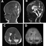
How is Germinal Matrix Hemorrhage Diagnosed?
Germinal matrix hemorrhage is diagnosed by ultrasound of the head and physical examination.
How is Germinal Matrix Hemorrhage Treated?
Germinal matrix hemorrhage is treated by antenatal dexamethasone administered to the mother or indomethacin administered to the infant. The ideal method is to prevent premature delivery.
What is the Prognosis of Germinal Matrix Hemorrhage?
The prognosis of germinal matrix hemorrhage is good for grade I and grade II germinal matrix hemorrhages. The prognosis depends on the extent of hemorrhage and the presence of hydrocephalus. Grades III and IV of germinal matrix hemorrhages have a poor prognosis. There is a 90% mortality for grade IV germinal matrix hemorrhages.