A mucocele is an oral lesion that is typically due to trauma.
What is the Pathology of Mucocele?
-The pathology of mucocele is:
-Etiology: The cause of mucocele is most often when a patient repeated trauma such as bites or sucking.
-Pathogenesis: The sequence of events that lead to mucoceles is the disruption of saliva flow from the salivary glands.
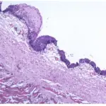
-Morphology: Small bump.
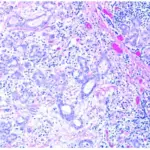
-Histology: The histology associated with mucocele shows intact pseudocyst cavity containing mucin, abundant epithelioid foamy histiocytes, neutrophils, and granulation tissue.
How does Mucocele Present?
Patients with mucoceles are typically younger, and are either male or female. The symptoms, features, and clinical findings associated with mucocele include painless, soft, round, dome-shaped, lesions that typically have pearly or clear or blue hues.
How is Mucocele Diagnosed?
Mucocele is diagnosed mainly based on the clinically by physical exam, and perhaps a biopsy.
How is Mucocele Treated?
Mucocele is treated by lifestyle changes, symptomatic management, or excisions.
What is the Prognosis of Mucocele?
The prognosis of mucocele is good.