Myocardial infarction is the irreversible death of heart muscle secondary to prolonged lack of oxygen supply. It typically occurs due to thromboembolism in a coronary artery.
What is the Pathology of Myocardial Infarction?
The pathology of myocardial infarction is:
-Etiology: The cause of myocardial infarction is coronary artery disease.
-Genes involved: None.
-Pathogenesis: The sequence of events that lead to myocardial infarction includes complete blockage of a coronary artery caused by a rupture of an atherosclerotic plaque
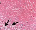
-Morphology: The morphology associated with myocardial infarction shows early morphologic indicators of irreversible myocardial damage are rupture of the sarcolemma and contraction band necrosis. The size of the infarction increases with the duration of ischemia.
-Histology: The histology associated with myocardial infarction shows left ventricle myocardium with the focus of coagulated necrosis, nuclei are localized in the center of cells.
How does Myocardial Infarction Present?
Patients with myocardial infarction typically affect males present at the age range of 30-64 years. The symptoms, features, and clinical findings associated with myocardial infarction include chest pain or discomfort which may travel into the shoulder, arm, back, neck or jaw, heartburn, shortness of breath, nausea, feeling faint, a cold sweat, or feeling tired.
How is Myocardial Infarction Diagnosed?
Myocardial infarction is diagnosed using ECG, blood tests (troponin), and coronary angiography.
How is Myocardial Infarction Treated?
Myocardial infarction is treated with aspirin, nitroglycerin, and opioids, supplemental oxygen, percutaneous coronary intervention, coronary artery bypass surgery, and angioplasty.
What is the Prognosis of Myocardial Infarction?
The prognosis of myocardial infarction is fair with a 30% mortality rate; about 50% of the deaths occur prior to arrival at the hospital. An additional 5-10% of survivors die within the first year after their myocardial infarction.