What is a Myofibroma?

Myofibroma
Myofibroma. (A–E) Clinical manifestations of hereditary leiomyomatosis and renal cell carcinoma (HLRCC): leiomyomas.Notes: (A) Multiple cutaneous leiomyomas on the torso of an HLRCC patient. (B) Histomicrograph of cutaneous leiomyoma, showing interlaced bundles of smooth-muscle fibers with a centrally located long, blunt-edged nucleus. (hematoxylin and eosin, [H&E] 10×). (C) Computed tomography image showing multiple large uterine leiomyomas (white arrows) that occur in most women with HLRCC, and that can be symptomatic. (D and E) High-power image of a uterine leiomyoma showing large prominent nuclei with an orangiophilic nucleolus (white arrows) surrounded by perinuclear halo; H&E, 300× and 400×. Images from Grubb et al32 (A and C), Rothman et al54 (B), and Sanz-Ortega et al30 (D and E). Hereditary leiomyomatosis and renal cell carcinoma. Schmidt LS, Linehan WM - International journal of nephrology and renovascular disease (2014). Not Altered. CC.

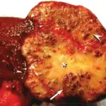
Myofibroma is a solitary nodular tumor of the soft tissue, bone, or internal organs.

What is the Pathology of Myofibroma? 

The pathology of Myofibroma is: 

-Etiology: The cause of myofibroma is a genetic mutation. 

-Genes involved: PDGFRB, SRF-RELA.

-Pathogenesis: The sequence of events that lead to myofibroma is the mutations in the PDGFRB gene.

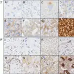
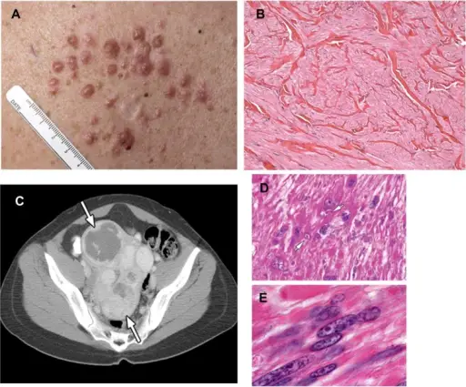
-Histology: The histology associated with myofibroma shows well-circumscribed, nodular neoplasms characterized by a distinctive biphasic growth pattern. 

How does Myofibroma Present?

Patients with myofibroma typically affect males in early childhood or adulthood present at the age range of 15 years and above. The symptoms, features, and clinical findings associated with myofibroma include a painless, slow-growing mass.

How is Myofibroma Diagnosed? 

Myofibroma is diagnosed by tissue sampling.

How is Myofibroma Treated? 

Myofibroma is treated by surgery.

What is the Prognosis of Myofibroma? 

The prognosis of myofibroma is good. They usually do not recur.