Odontogenic keratocyst is the cyst arising from the cell rests of the dental lamina. It can occur anywhere in the jaw but is commonly seen in the posterior part of the mandible.
What is the Pathology of Odontogenic Keratocyst?
The pathology of odontogenic keratocyst is:
-Etiology: The cause of odontogenic keratocyst is generally thought to be derived from remnants of the dental lamina.
-Pathogenesis: The sequence of events that lead to odontogenic keratocyst is two-hit mechanism results in bi-allelic loss of PTCH (“patched”), tumor suppressor, on 9q22.3-q31 causing dysregulation of p53 and cyclin D1 oncoproteins.
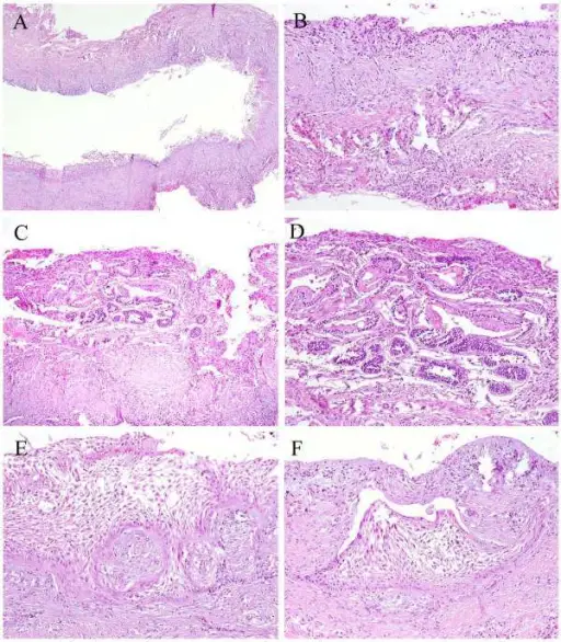
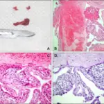
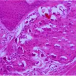
-Histology: The histology associated with odontogenic keratocyst shows uniform epithelial lining 6 – 8 cells thick lacking rete ridges.
How does Odontogenic Keratocyst Present?
Patients with odontogenic keratocyst typically affect males and females present in the age range of 30-60 years. The symptoms, features, and clinical findings associated with odontogenic keratocyst include bony expansion or infection. However, bony expansion is uncommon as odontogenic keratocysts grow due to increased epithelial turnover rather than osmotic pressure. When symptoms are present they usually take the form of pain, swelling, and discharge due to secondary infection.
How is Odontogenic Keratocyst Diagnosed?
Odontogenic keratocyst is diagnosed usually radiologically. However, a definitive diagnosis is through biopsy.
How is Odontogenic Keratocyst Treated?
Odontogenic keratocysts are treated by resection or enucleation.
What is the Prognosis of Odontogenic Keratocyst?
The prognosis of odontogenic keratocyst is poor. Recurrent OKC may develop in three different ways: By incomplete removal of the original cyst lining; by the retention of daughter cysts, from microcysts or epithelial islands in the wall of the original cyst, or by the development of new OKC from epithelial off-shoots of the basal layer.


