A pulmonary embolism is a blockage in one of the pulmonary arteries in the lungs.
What is the Pathology of Pulmonary Embolism?
The pathology of pulmonary embolism is:
-Etiology: The cause of pulmonary embolism is a clump of material that gets wedged into an artery in the lungs, such as blood clots, fat from the marrow of a broken long bone, part of a tumor, and air bubbles. Pulmonary embolisms can be caused by blood clots that travel to the lungs from deep veins in the legs. Deep vein thrombosis is a rare condition in which blood clots travel from veins in other parts of the body.
-Pathogenesis: The sequence of events that lead to pulmonary embolism is the portions of the lung with blocked arteries that are robbed of blood and may die.
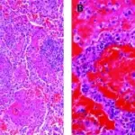
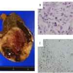
-Morphology: The morphology associated with pulmonary embolism shows multiple clots in many cases, obstructions in the arteries inside your lungs, and pulmonary infarction.
How does Pulmonary Embolism Present?
Patients with pulmonary embolism typically are either males or females present at the age range of older years. The symptoms, features, and clinical findings associated with pulmonary embolism include shortness of breath, chest pain that’s felt when breathing in deeply, coughing, bending or stooping, and cough. Other signs and symptoms that can occur include rapid or irregular heartbeat, lightheadedness or dizziness, fever, excessive sweating, and clammy or discolored skin (cyanosis). Leg pain and swelling are caused by a deep vein thrombosis and usually occur in the calf. Pulmonary embolism can also lead to pulmonary hypertension.
How is Pulmonary Embolism Diagnosed?
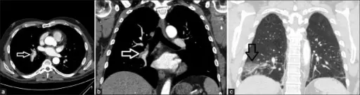
Pulmonary embolism is diagnosed with a medical history, a physical exam, and tests that include blood tests, chest x-ray, CT pulmonary angiography, ultrasound, MRI, ventilation-perfusion scan (V/Q scan), and pulmonary angiogram.
How is Pulmonary Embolism Treated?
Pulmonary embolism is treated with medications, such as blood thinners (anticoagulants) and clot dissolvers (thrombolytics). Surgical procedures include clot removal and vein filter. It’s important to continue treatment because of the risk of another deep vein thrombosis or pulmonary embolism.
What is the Prognosis of Pulmonary Embolism?
The prognosis of pulmonary embolism is fair. It can be life-threatening, but prompt treatment greatly reduces the risk of death. Protection against pulmonary embolism includes taking measures to prevent blood clots in the legs.