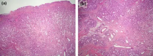
Under an ulcerated surface, there is an extensive granulation tissue reaction characterized by abundant small size vessels arranged perpendicularly to the surface. At the bottom of the image, the characteristic appearance of pyogenic granuloma can be seen on the right (H&E, original magnification 10x). (b). At higher power, the typical histologic features of pyogenic granuloma consist in a proliferation of capillary-sized vessels arranged surrounding larger vessel structures producing its typical lobular distribution. There is stromal fibrosis dissecting between the capillary lobules. (H&E, original magnification 20x). A Gray-purple Mass on the Floor of the Mouth: Gigantic Mucogingival Pyogenic Granuloma in a Teenage Patient: Brunet-LLobet L, Miranda-Rius J, Lahor-Soler E, Mrina O, Nadal A - The open dentistry journal (2014). Not altered. CC.
A pyogenic granuloma is a benign overgrowth of capillaries. Pyogenic granulomas are also referred to as capillary granulomas or lobular capillary hemangiomas. They are often found on the gums, nasal septum, and skin. They may occur at any age.
Pathology
Cause: Pyogenic granulomas are due to proliferation of capillaries.
Gross: Red fleshy nodule.
Microscopic: Lobular vascular proliferation.
Stains: No special stains needed.
Differential Diagnosis: Acrodermatitis, Bacillary Angiomatosis, Infantile hemangioendothelioma, Verruga peruana.
Presentation
Red dome shaped bump.
Diagnosis
Made histopathologically.
Treatment
Pyogenic granulomas may be surgically removed.
Prognosis
Great. Pyogenic granulomas may regress, develop into dense fibrous masses, or turn into peripheral ossifying fibromas.


