A Rathke cleft cyst is a benign fluid-filled lesion that develops between the structure of the anterior pituitary gland at the base of the brain hypothesized to instigate from fragments of Rathke pouch.
What is the Pathology of Rathke Cleft Cyst?
The pathology of Rathke cleft cyst is:
-Etiology: The cause of Rathke cleft cyst is the failure of Rathke cleft to regress.
-Genes involved: FOXA1, and FOXJ1.
-Pathogenesis: The sequence of events that lead to Rathke cleft cyst, resulting from true fragments of the embryologic Rathke pouch, the pouch residual lumen reduced to a thin Rathke cleft. This cleft is supposed to regress. Its persistence leads to Rathke cleft cyst.
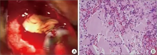
-Morphology: The morphology associated with Rathke cleft cyst shows thin presenting with different colors, (blue, yellow, gray, white, pink, red, or green). Varying in size 10 and 20mm, gelatinous yellow fluid.
-Histology: The histology associated with rathke cleft cyst shows vascularized stroma of connective tissue, mucous secreting ciliated, and non-ciliated epithelial cells.
How does Rathke Cleft Cyst Present?
Patients with Rathke cleft cyst typically have a 2:1 female to male ratio present at age range 30-50 years of age. The symptoms, features, and clinical findings associated with Rathke cleft cyst include chiasmatic syndrome, vision changes, headaches, nausea, fatigue, rare epilepsy, symptoms of hypopituitarism, growth retardation. low libido, and impotence.
How is Rathke Cleft Cyst Diagnosed?
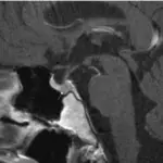
Rathke cleft cyst is diagnosed through radiography, plain skull indicates abnormal configured sellar, CT scan and brain MRI.
How is Rathke Cleft Cyst Treated?
Rathke cleft cyst is treated through transsphenoidal surgery.
What is the Prognosis of Rathke Cleft Cyst?
The prognosis of a rathke cleft cyst is good.


