Thymic carcinomas are rare types of aggressive cancers of thymic epithelial origin.
What is Thymic Carcinoma?

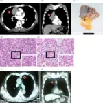
a Macroscopic findings of cross-sectional slices showed a small solid component (8 mm). b Microscopy revealed a proliferation of markedly atypical polygonal epithelial cells having hyperchromatic nuclei (×400). c Immunohistochemically, tumor cells are positive for CD5 (×400). Middle mediastinal thymic carcinoma with cystic findings in radiologic images: a case report: Surgical Case Reports. Not altered. CC.


