Acute necrotizing hemorrhagic encephalomyelitis is a rare, demyelinating disease of the CNS characterized by rapidly progressive inflammation of the white matter. It is the most severe form of acute disseminated encephalomyelitis.
What is Acute Necrotizing Hemorrhagic Encephalomyelitis?

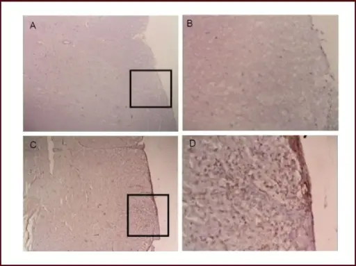
PD-L1 immunoreactivity in the spinal cord of experimental allergic encephalomyelitis mice.(A, B) Control group (A: × 100, B: × 400). (C, D) Model group at 2 days after onset (C: × 100, D: × 400). (B, D) Higher magnification views of boxed areas in A, C. In the experimental allergic encephalomyelitis mice, brown staining on infiltrating inflammatory cells indicated PD-L1 expression. In the control group, no inflammatory cells were present and PD-L1 molecule immunoreactivity was not observed.PD-L1 is increased in the spinal cord and infiltrating lymphocytes in experimental allergic encephalomyelitis.
Li M, Jiang J, Fu B, Chen J, Xue Q, Dong W, Gu Y, Tang L, Xue L, Fang Q, Wang M, Zhang X - Neural regeneration research (2013). Not Altered. CC


