Age related macular degeneration is the loss of central vision induced by the degeneration of the macular of the retina manifesting after age 50.
What is the Pathology of Age Related Macular Degeneration?
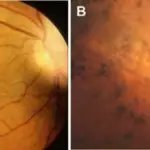
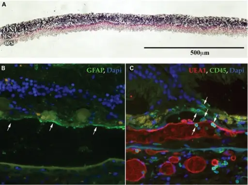
The pathology of age related macular degeneration is degeneration involving the retinal photoreceptors, pigment epithelium and Bruch’s membrane.
How does Age Related Macular Degeneration Present?
Age related macular degeneration presents with blurry vision, distorted vision, partial vision loss, and neovascularization.
How is Age Related Macular Degeneration Diagnosed?
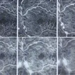
Age related macular degeneration is diagnosed by Amsler grid test, pupillary dilation test, optical coherence tomography, and fluorescein angiography.
How is Age Related Macular Degeneration Treated?
Age related macular degeneration is treated with anti-VEGF medications, vitamin supplementation, and laser therapy.
What is the Prognosis of Age Related Macular Degeneration?
The prognosis of age related macular degeneration can last for years or life-long.