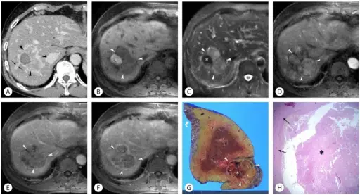
(A). Axial T1-weighted fat-suppressed MR image shows that a 6 cm lobulated mass (arrowheads) in the right hepatic lobe, which consists of large area of hypointensity and small area of hyperintensity (asterisk) (B). Axial T2-weighted fat-suppressed MR image shows that a mass (arrowheads) in the right hepatic lobe consists of large area of hyperintensity and small area of hypointensity (asterisk), which looks reversed with respect to signal intensity on B (C). Axial gadolinium-enhanced T1-weighted arterial (D), portal (E), and delayed-phase (F) MR images show early arterial enhancement of the tumor that is accompanied by portal and delayed wash-out (arrowheads) with overally mosaic pattern of enhancement. And also, well defined tumoral capsule with delayed enhancement is seen on F (D-F). Photograph of gross specimen shows a 6 cm brown to reddish lobulated solid and cystic mass with surrounding whitish fibrous capsule (arrowheads), hemorrhagic necrosis (asterisk), necrotic debris and internal fibrous septa (arrows) within the resected right hepatic lobe. Note focal non-smooth surface (curved arrow) of the liver, which is suggestive of chronic alcoholic liver disease (G). Photomicrograph shows a necrotic lesion (asterisk) with surrounding fibrous capsule (arrows), which consists of extensive areas of coagulative necrosis, infiltrations of chronic inflammatory cells including lymphoplasma cells and some eosinophils, and fibrosis (H) (Hematoxylin and Eosin stain, ×40). Hepatic abscess mimicking hepatocellular carcinoma in a patient with alcoholic liver disease: Kim JW, Shin SS, Heo SH, Lim HS, Hur YH, Kim JH - Clinical and molecular hepatology (2013). Not altered. CC.
Alcoholic liver disease is a result of over-consuming alcohol that damages the liver, leading to a buildup of fats, inflammation, and scarring.
What is the Pathology of Alcoholic Liver Disease?
The pathology of alcoholic liver disease is:
-Etiology: The cause of the alcoholic liver disease is overconsuming alcohol.
-Genes involved: None.
-Pathogenesis: The sequence of events that lead to an alcoholic liver disease involves secretion of pro-inflammatory, oxidative stress, lipid peroxidation, and acetaldehyde toxicity. These factors cause inflammation, apoptosis, and eventually fibrosis of liver cells.
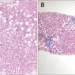
-Histology: The histology associated with alcoholic liver disease shows macrovesicular steatosis, neutrophilic lobular inflammation, ballooning degeneration, Mallory-Denk bodies, portal, and fibrosis that is pericellular.
How does Alcoholic Liver Disease Present?
Patients with the alcoholic liver disease typically affect females present in the age range of 25-34. The symptoms, features, and clinical findings associated with alcoholic liver disease include abdominal pain and tenderness, dry mouth and increased thirst, fatigue, jaundice, portal hypertension, enlarged spleen, ascites, and kidney failure.
How is Alcoholic Liver Disease Diagnosed?
Alcoholic liver disease is diagnosed by blood tests, liver biopsy, CT scan, and ultrasound.
How is Alcoholic Liver Disease Treated?
Alcoholic liver disease is treated medical and lifestyle management.
What is the Prognosis of Alcoholic Liver Disease?
The prognosis of alcoholic liver disease is fair with a survival rate of 60%.


