An infarction is an ischemic necrosis due to occlusion of venous drainage or arterial supply.
What is an Infarction?

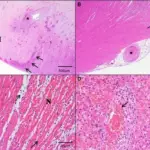
Infarction. Microscopic slides at 200× magnification after standard hematoxylin and eosin staining of three samples of the heart of a 44-year-old man who died 3 days after admission with acute inferoposterior infarction (patient not included in the study group). (a) infarct core with necrotic cardiomyocytes (interrupted arrow) and abundant erythrocytes (solid arrows), (b) infarct border zone with edema (open arrow), (c) capillary vessel with thrombus (asterisk) and plugged polymorphonuclear cells (circled arrow). Intramyocardial hemorrhage and microvascular obstruction after primary percutaneous coronary intervention. Beek AM, Nijveldt R, van Rossum AC - The international journal of cardiovascular imaging (2009). Not Altered. CC.


