Osteoid osteoma is a benign bone tumor that arises from osteoblasts and some components of osteoclasts.
What is the Pathology of Osteoid Osteoma?
The pathology of osteoid osteoma is:
-Etiology: The cause of osteoid osteoma remains uncertain.
-Genes involved: YWHAH and PDGF-B.
-Pathogenesis: The sequence of events that lead to osteoid osteoma occurs when certain cells divide uncontrollably, forming a small mass of bone and other tissue.
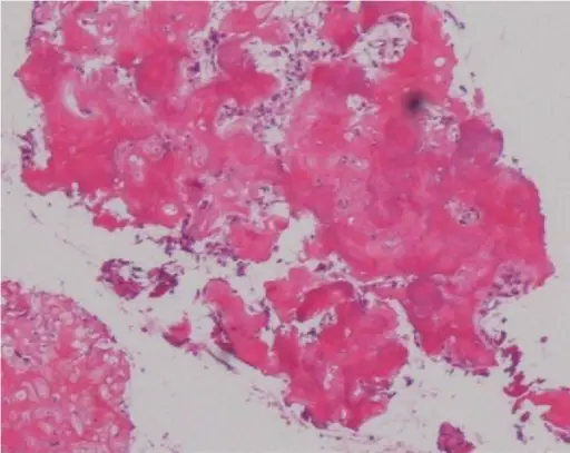
-Histology: The histology associated with osteoid osteoma shows small, yellow to the red nidus of osteoid and woven bone with interconnected trabeculae, and background and rim of highly vascularized, fibrous connective tissue.
How does Osteoid Osteoma Present?
Patients with osteoid osteoma typically affect males 3 times more than females in 20-30 years of age. The symptoms, features, and clinical findings associated with osteoid osteoma include dull pain that escalates to severe at night or slight pain, rising to become severe even at night-time, affecting sleep quality, limping, muscle atrophy, bowing deformity, swelling, increased or decreased bone growth.
How is Osteoid Osteoma Diagnosed?
Osteoid osteoma is diagnosed by radiographs.
How is Osteoid Osteoma Treated?
Osteoid osteoma is treated by conservative management with NSAIDs.
What is the Prognosis of Osteoid Osteoma?
The prognosis of osteoid osteoma is good since it is a benign process with no potential for malignant degeneration.


