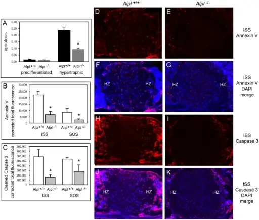
(A) Undifferentiated rib chondrocytes and differentiated rib hypertrophic chondrocytes from Alpl+/+ and Alpl−/− mice were cultured in media containing 10% FBS. The generation of apoptotic changes in DNA was assayed by a colorimetric reaction. Results show no difference in the tendency for pre-differentiated cells to undergo apoptosis and a significantly diminished tendency for differentiated hypertrophic chondrocytes to undergo apoptosis. (B-K) ISS and SOS cranial base tissues were stained for Annexin V and cleaved Caspase 3 markers of apoptosis by immunofluorescence. Images show diminished expression of Annexin V in Alpl−/−(E, G) compared to Alpl+/+(D, F) ISS, and diminished expression of cleaved Caspase 3 in Alpl−/−(I, K) compared to Alpl+/+(H, J) ISS. Tissue Nonspecific Alkaline Phosphatase (TNAP) Regulates Cranial Base Growth and Synchondrosis Maturation: Frontiers in Physiology. Not altered. CC.
Apoptosis is programmed cell death.
Causes of apoptosis are cellular stressors which include ischemia, hypoxia, infectious agents, radiation, chemicals, alterations in temperature.
Apoptosis in Physiologic Situations: Natural.
Apoptosis in Pathologic Conditions: Disease or disorder.
Morphologic and Biochemical Changes in Apoptosis: Homeostasis disrupted.
Mechanisms of Apoptosis: Intrinsic and Extrinsic.
The Intrinsic (Mitochondrial) Pathway of Apoptosis
The Extrinsic (Death Receptor-Initiated) Pathway of Apoptosis
The Execution Phase of Apoptosis
Dead Cell Removal
Necroptosis