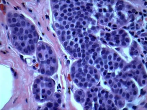
Atypical lobular hyperplasia is a clonal proliferation of discohesive epithelial cells arising in terminal duct lobular units.
What is the Pathology of Atypical Lobular Hyperplasia?
The pathology of atypical lobular hyperplasia is:
-Etiology: The cause of atypical lobular hyperplasia is the increased expression of ESR1 and decreased expression of SFRP1.
-Pathogenesis: The sequence of events that lead to atypical lobular hyperplasia is the 16q loss or CDH1 deletion and loss of function of E-cadherin.
-Morphology: The morphology associated with atypical lobular hyperplasia shows loosely cohesive cell clusters composed of uniform cells with occasional intracytoplasmic lumina with minimal nuclear atypia.
-Histology: The histology associated with atypical lobular hyperplasia shows the solid, discohesive proliferation of cells that are monomorphic, small, have pale pink cytoplasm, uniform oval nuclei, and indistinct nuclei.
How does Atypical Lobular Hyperplasia Present?
Patients with atypical lobular hyperplasia typically are more female present at age range of wide age groups, but most commonly diagnosed in 50s.
How is Atypical Lobular Hyperplasia Diagnosed?
Atypical lobular hyperplasia is diagnosed by core biopsy, mammography, ultrasound, or MRI.
How is Atypical Lobular Hyperplasia Treated?
Atypical lobular hyperplasia is treated with selective estrogen receptor modulators (SERMs) and aromatase inhibitors (AIs).
What is the Prognosis of Atypical Lobular Hyperplasia?
The prognosis of atypical lobular hyperplasia is fair.


